https://doi.org/10.1140/epje/s10189-025-00497-0
Regular Article - Living Systems
Osmotic pressure induces unexpected relaxation of contractile 3D microtissue
1
Laboratoire Interdisciplinaire de Physique (LIPhy), Université Grenoble Alpes, CNRS, 38000, Grenoble, France
2
Université Paris Cité, CNRS, Institut Jacques Monod, 75013, Paris, France
3
Institut de Biologie Paris-Seine (IBPS), Sorbonne Université, CNRS, 75005, Paris, France
4
Department of Physics, University of Erlangen-Nürnberg, Erlangen, Germany
5
Dipartimento Di Scienze Matematiche, Politecnico Di Torino, Turin, Italy
a
giovanni.cappello@univ-grenoble-alpes.fr
b
thomas.boudou@cnrs.fr
Received:
25
February
2025
Accepted:
28
May
2025
Published online:
24
June
2025
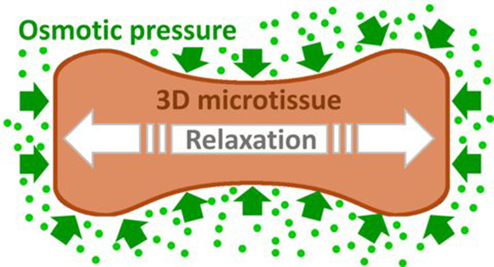
Cell contraction and proliferation, matrix secretion and external mechanical forces induce compression during embryogenesis and tumor growth, which in turn regulate cell proliferation, metabolism or differentiation. How compression affects tissue contractility, a hallmark of tissue function, is however unknown. Here we apply osmotic compression to microtissues of either mouse colon adenocarcinoma CT26 cells, mouse NIH 3T3 fibroblasts, or human primary colon cancer-associated fibroblasts. Microtissues are anchored to flexible pillars that serve as force transducers. We observe that low-amplitude osmotic compression induces a rapid relaxation of tissue contractility, primed by the deformation of the extracellular matrix. Furthermore, we show that this compression-induced relaxation is independent of the cell type, proportional to the initial tissue contractility, and depends on RhoA-mediated myosin activity. Together, our results demonstrate that compressive stress can relax active tissue force, and points to a potential role of this feedback mechanism during morphogenetic events such as onco- or embryogenesis.
Supplementary Information The online version contains supplementary material available at https://doi.org/10.1140/epje/s10189-025-00497-0.
© The Author(s) 2025
 Open Access This article is licensed under a Creative Commons Attribution 4.0 International License, which permits use, sharing, adaptation, distribution and reproduction in any medium or format, as long as you give appropriate credit to the original author(s) and the source, provide a link to the Creative Commons licence, and indicate if changes were made. The images or other third party material in this article are included in the article's Creative Commons licence, unless indicated otherwise in a credit line to the material. If material is not included in the article's Creative Commons licence and your intended use is not permitted by statutory regulation or exceeds the permitted use, you will need to obtain permission directly from the copyright holder. To view a copy of this licence, visit http://creativecommons.org/licenses/by/4.0/.