https://doi.org/10.1140/epje/s10189-023-00404-5
Regular Article - Living Systems
Vesicle condensation induced by synapsin: condensate size, geometry, and vesicle shape deformations
1
Institut für Röntgenphysik, Georg-August-Universität, Friedrich-Hund-Platz 1, 37077, Göttingen, Germany
2
Molekulare Neurowissenschaften, Deutsches Zentrum für Neurodegenerative Erkrankungen (DZNE), Charitéplatz 1, 10117, Berlin, Germany
3
Labor für Neurobiologie, Max-Planck-Institut für multidisziplinäre Naturwissenschaften, Am Fassberg 11, 37077, Göttingen, Germany
4
Institut für Neuropathologie, Universitätsmedizin Göttingen, Justus-von-Liebig-Weg 11, 37077, Göttingen, Germany
5
Theorie Biologischer Flüssigkeiten, Max-Planck-Institut für Dynamik und Selbstorganisation, Am Fassberg 11, 37077, Göttingen, Germany
Received:
26
July
2023
Accepted:
28
December
2023
Published online:
25
January
2024
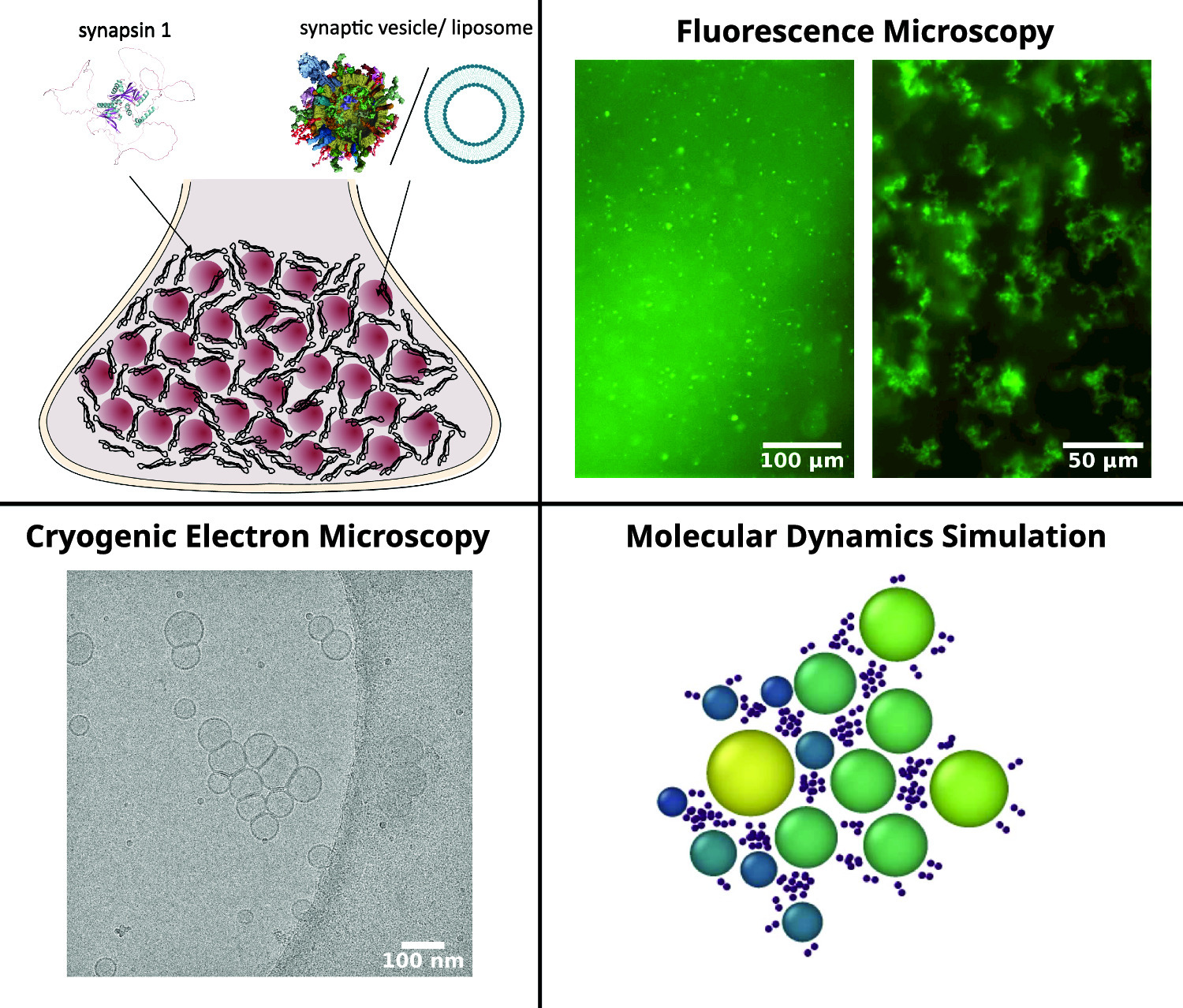
We study the formation of vesicle condensates induced by the protein synapsin, as a cell-free model system mimicking vesicle pool formation in the synapse. The system can be considered as an example of liquid–liquid phase separation (LLPS) in biomolecular fluids, where one phase is a complex fluid itself consisting of vesicles and a protein network. We address the pertinent question why the LLPS is self-limiting and stops at a certain size, i.e., why macroscopic phase separation is prevented. Using fluorescence light microscopy, we observe different morphologies of the condensates (aggregates) depending on the protein-to-lipid ratio. Cryogenic electron microscopy then allows us to resolve individual vesicle positions and shapes in a condensate and notably the size and geometry of adhesion zones between vesicles. We hypothesize that the membrane tension induced by already formed adhesion zones then in turn limits the capability of vesicles to bind additional vesicles, resulting in a finite condensate size. In a simple numerical toy model we show that this effect can be accounted for by redistribution of effective binding particles on the vesicle surface, accounting for the synapsin-induced adhesion zone.
Jette Alfken and Charlotte Neuhaus have contributed equally to this work.
Supplementary Information The online version contains supplementary material available at https://doi.org/10.1140/epje/s10189-023-00404-5.
© The Author(s) 2024
 Open Access This article is licensed under a Creative Commons Attribution 4.0 International License, which permits use, sharing, adaptation, distribution and reproduction in any medium or format, as long as you give appropriate credit to the original author(s) and the source, provide a link to the Creative Commons licence, and indicate if changes were made. The images or other third party material in this article are included in the article’s Creative Commons licence, unless indicated otherwise in a credit line to the material. If material is not included in the article’s Creative Commons licence and your intended use is not permitted by statutory regulation or exceeds the permitted use, you will need to obtain permission directly from the copyright holder. To view a copy of this licence, visit http://creativecommons.org/licenses/by/4.0/.